web 2.0

Saturday, June 26, 2010

Top 10 Deadliest Diseases

One of the most tragic parts of being human is being afflicted by diseases with high morbidity and mortality. Here is our list of top 10 deadliest diseases ever known to man.
aids1.    AIDS; 25 million from 1981 to present
AIDS is a mix of infections and complications as a result of progressive damage to the body’s immune system caused by HIV. AIDS is now considered a pandemic.

influenza2.    Influenza; 36,000 deaths annually
Influenza, which is more commonly known as flu, is a highly infectious disease that is caused by influenza virus. Transmission of the disease is by airborne and through physical contact.

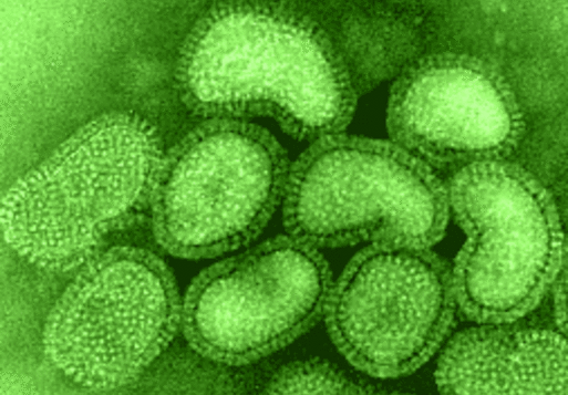
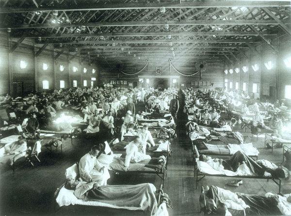
spanish_flu3.    Spanish Flu, 1918-19; 100 million deaths
The flu pandemic that happened in 1918 is termed as a category 5 flu pandemic which was caused by the flu virus strain A with subtype H1N1.


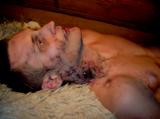
bubonic-plague4.    Bubonic Plague; 250 million deaths

This disease outbreak was mainly caused by fleas and rodents infected with Xenopsylla cheopsis. Humans were infected after being bitten by an affected rodent.

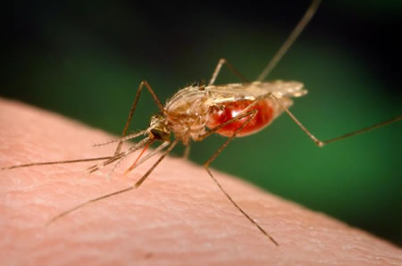
malaria5.    Malaria; 2.7 million deaths annually
Malaria is an infectious disease which is vector-borne. The causative agent is the protozoan parasites. It is a common disease in the sub-tropics and tropical regions.


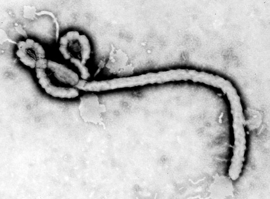
ebola6.    Ebola; 160,000 deaths from 2000 to present

The Ebola virus was first isolated in1976 from the dual outbreaks that occurred in Zaire and Sudan. It is a zoonotic disease as it affects lowland apes as well as humans.

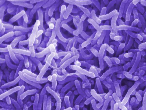
cholera7.    Cholera; 12,000 deaths from 1991 to present
The epidemic or Asiatic cholera is a very serious type of diarrheal ailment caused by Vibrio cholera. The mode of transmission is by ingesting contaminated food and water.

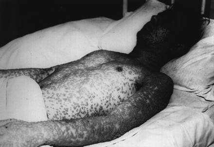
smallpox8.    Smallpox; Population drop from 12 million to 235,000
Smallpox is a highly contagious viral disease that has two variants. The V major has a 35% mortality rate while the less severe V minor has a 1% mortality rate.

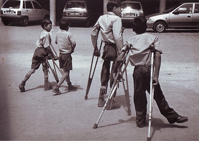
polio9.    Polio, 10,000 deaths from 1916 to present
Polio or infantile paralysis is a viral disease that is transmitted through the fecal-oral course.


black-death10.
Black Death; 75 million deaths
The Black Plague is the one of the most serious pandemic outbreak in modern history. It was believed to have started in Central Asia and affected Europe during the 13th century.

Thursday, June 17, 2010

Hemochromatosis

What is hemochromatosis?

Hemochromatosis is the most common form of iron overload disease. Primary hemochromatosis, also called hereditary hemochromatosis, is an inherited disease. Secondary hemochromatosis is caused by anemia, alcoholism, and other disorders.
Juvenile hemochromatosis and neonatal hemochromatosis are two additional forms of the disease. Juvenile hemochromatosis leads to severe iron overload and liver and heart disease in adolescents and young adults between the ages of 15 and 30. The neonatal form causes rapid iron buildup in a baby’s liver that can lead to death.
Illustration of the digestive system with heart, liver, and pancreas highlighted.
Excess iron is stored in body tissues, specifically the liver, heart, and pancreas.
Hemochromatosis causes the body to absorb and store too much iron. The extra iron builds up in the body’s organs and damages them. Without treatment, the disease can cause the liver, heart, and pancreas to fail.
Iron is an essential nutrient found in many foods. The greatest amount is found in red meat and iron-fortified breads and cereals. In the body, iron becomes part of hemoglobin, a molecule in the blood that transports oxygen from the lungs to all body tissues.
Healthy people usually absorb about 10 percent of the iron contained in the food they eat, which meets normal dietary requirements. People with hemochromatosis absorb up to 30 percent of iron. Over time, they absorb and retain between five to 20 times more iron than the body needs.
Because the body has no natural way to rid itself of the excess iron, it is stored in body tissues, specifically the liver, heart, and pancreas.

What causes hemochromatosis?

Hereditary hemochromatosis is mainly caused by a defect in a gene called HFE, which helps regulate the amount of iron absorbed from food. The two known mutations of HFE are C282Y and H63D. C282Y is the most important. In people who inherit C282Y from both parents, the body absorbs too much iron and hemochromatosis can result. Those who inherit the defective gene from only one parent are carriers for the disease but usually do not develop it; however, they still may have higher than average iron absorption. Neither juvenile hemochromatosis nor neonatal hemochromatosis are caused by an HFE defect. Juvenile and neonatal hemochromatosis are caused by a mutation in a gene called hemojuvelin.


What are the risk factors of hemochromatosis?

Hereditary hemochromatosis is one of the most common genetic disorders in the United States. It most often affects Caucasians of Northern European descent, although other ethnic groups are also affected. About five people out of 1,000—0.5 percent—of the U.S. Caucasian population carry two copies of the hemochromatosis gene and are susceptible to developing the disease. One out of every 8 to 12 people is a carrier of one abnormal gene. Hemochromatosis is less common in African Americans, Asian Americans, Hispanics/Latinos, and American Indians.
Although both men and women can inherit the gene defect, men are more likely than women to be diagnosed with hereditary hemochromatosis at a younger age. On average, men develop symptoms and are diagnosed between 30 to 50 years of age. For women, the average age of diagnosis is about 50.

What are the symptoms of hemochromatosis?

Joint pain is the most common complaint of people with hemochromatosis. Other common symptoms include fatigue, lack of energy, abdominal pain, loss of sex drive, and heart problems. However, many people have no symptoms when they are diagnosed.
If the disease is not detected and treated early, iron may accumulate in body tissues and eventually lead to serious problems such as
  • arthritis
  • liver disease, including an enlarged liver, cirrhosis, cancer, and liver failure
  • damage to the pancreas, possibly causing diabetes
  • heart abnormalities, such as irregular heart rhythms or congestive heart failure
  • impotence
  • early menopause
  • abnormal pigmentation of the skin, making it look gray or bronze
  • thyroid deficiency
  • damage to the adrenal glands

How is hemochromatosis diagnosed?

A thorough medical history, physical examination, and routine blood tests help rule out other conditions that could be causing the symptoms. This information often provides helpful clues, such as a family history of arthritis or unexplained liver disease.
  • Blood tests can determine whether the amount of iron stored in the body is too high. The transferrin saturation test reveals how much iron is bound to the protein that carries iron in the blood. Transferrin saturation values higher than 45 percent are considered too high.
  • The total iron binding capacity test measures how well your blood can transport iron, and the serum ferritin test shows the level of iron in the liver. If either of these tests shows higher than normal levels of iron in the body, doctors can order a special blood test to detect the HFE mutation, which will confirm the diagnosis. If the mutation is not present, hereditary hemochromatosis is not the reason for the iron buildup and the doctor will look for other causes.
  • A liver biopsy may be needed, in which case a tiny piece of liver tissue is removed and examined with a microscope. The biopsy will show how much iron has accumulated in the liver and whether the liver is damaged.
Hemochromatosis is considered rare and doctors may not think to test for it. Thus, the disease is often not diagnosed or treated. The initial symptoms can be diverse, vague, and mimic the symptoms of many other diseases. The doctors also may focus on the conditions caused by hemochromatosis—arthritis, liver disease, heart disease, or diabetes—rather than on the underlying iron overload. However, if the iron overload caused by hemochromatosis is diagnosed and treated before organ damage has occurred, a person can live a normal, healthy life.
Hemochromatosis is usually treated by a specialist in liver disorders called a hepatologist, a specialist in digestive disorders called a gastroenterologist, or a specialist in blood disorders called a hematologist. Because of the other problems associated with hemochromatosis, other specialists may be involved in treatment, such as an endocrinologist, cardiologist, or rheumatologist. Internists or family practitioners can also treat the disease.

How is hemochromatosis treated?

Treatment is simple, inexpensive, and safe. The first step is to rid the body of excess iron. This process is called phlebotomy, which means removing blood the same way it is drawn from donors at blood banks. Based on the severity of the iron overload, a pint of blood will be taken once or twice a week for several months to a year, and occasionally longer. Blood ferritin levels will be tested periodically to monitor iron levels. The goal is to bring blood ferritin levels to the low end of normal and keep them there. Depending on the lab, that means 25 to 50 micrograms of ferritin per liter of serum.
Once iron levels return to normal, maintenance therapy begins, which involves giving a pint of blood every 2 to 4 months for life. Some people may need phlebotomies more often. An annual blood ferritin test will help determine how often blood should be removed. Regular follow-up with a specialist is also necessary.
If treatment begins before organs are damaged, associated conditions—such as liver disease, heart disease, arthritis, and diabetes—can be prevented. The outlook for people who already have these conditions at diagnosis depends on the degree of organ damage. For example, treating hemochromatosis can stop the progression of liver disease in its early stages, which leads to a normal life expectancy. However, if cirrhosis, or scarring of the liver, has developed, the person’s risk of developing liver cancer increases, even if iron stores are reduced to normal levels.
People with complications of hemochromatosis may want to receive treatment from a specialized hemochromatosis center. These centers are located throughout the country.
People with hemochromatosis should not take iron or vitamin C supplements. And those who have liver damage should not consume alcoholic beverages or raw seafood because they may further damage the liver.
Treatment cannot cure the conditions associated with established hemochromatosis, but it will help most of them improve. The main exception is arthritis, which does not improve even after excess iron is removed.

How is hemochromatosis tested?

Screening for hemochromatosis—testing people who have no symptoms—is not a routine part of medical care or checkups. However, researchers and public health officials do have some suggestions.
  • Siblings of people who have hemochromatosis should have their blood tested to see if they have the disease or are carriers.
  • Parents, children, and other close relatives of people who have the disease should consider being tested.
  • Doctors should consider testing people who have joint disease, severe and continuing fatigue, heart disease, elevated liver enzymes, impotence, and diabetes because these conditions may result from hemochromatosis.
Since the genetic defect is common and early detection and treatment are so effective, some researchers and education and advocacy groups have suggested that widespread screening for hemochromatosis would be cost-effective and should be conducted. However, a simple, inexpensive, and accurate test for routine screening does not yet exist and the available options have limitations. For example, the genetic test provides a definitive diagnosis, but it is expensive. The blood test for transferrin saturation is widely available and relatively inexpensive, but it may have to be done twice with careful handling to confirm a diagnosis and show that the result is the consequence of iron overload.

Hope through Research

Scientists hope further study of the HFE gene will reveal how the body normally metabolizes iron. They also want to learn how iron injures cells and contributes to organ damage in other diseases, such as alcoholic liver disease, hepatitis C, porphyria cutanea tarda, heart disease, reproductive disorders, cancer, autoimmune hepatitis, diabetes, and joint disease.
Scientists are working to find out why only some patients with HFE mutations develop the disease. In addition, hemochromatosis research includes the following areas:
Genetics. Researchers are examining how the HFE gene normally regulates iron levels and why not everyone with an abnormal pair of genes develops the disease.
Pathogenesis. Scientists are studying how iron injures body cells. Iron is an essential nutrient, but above a certain level it can damage or even kill cells.
Epidemiology. Research is underway to explain why the amounts of iron people normally store in their bodies differ. Research is also being conducted to determine how many people with the defective HFE gene go on to develop symptoms and why some people develop symptoms and others do not.
Screening and testing. Scientists are working to determine at what age testing is most effective, which groups should be tested, and which are the best tests for widespread screening.

Mutant sperm guide clinicians to new diseases

Disease, DNA, deletions and duplications in human sperm

Human sperm
Human sperm:
Research published today in Nature Genetics shows that some rearrangements of the human genome occur more frequently than previously thought. The work is likely to lead to new identification of genes involved in disease and to improve diagnosis of genomic disease.
The scientists from the Wellcome Trust Sanger Institute looked at four unstable regions in the genome where rearrangements cause genetic diseases, so-called 'genomic disorders', and found that some of these rearrangements were found in sperm much more frequently than expected.
The team, led by Dr Matt Hurles, showed that losses or duplication of 'chunks' of the human genome occurred frequently in apparently healthy people. These losses or gains of DNA regions are called Copy Number Variants (CNVs), and can be found all over the genome in every individual.
Some of the mechanisms thought to produce CNVs would be expected to produce about one duplication for every deletion: however, clinical records for genomic disorders show only a few duplications, compared with hundreds of deletions.
" These are unfortunate accidents of the essential shuffling of our genetic deck of cards, a process essential to human life. "
Dr Matt Hurles
"There was no direct, global measure of the relative rate at which human DNA is gained or lost, a study that requires many thousands of human genomes," explained Dr Matt Hurles, Investigator at the Wellcome Trust Sanger Institute, "so we carried out a study on four clinically important regions using human sperm cells as our population of genomes. 
"Sperm cells give us an unbiased snapshot of CNVs: using our new highly-sensitive assays we can detect one rearrangement in a million cells."
The team looked at regions known to be affected by rearrangement in Williams-Beuren Syndrome, Charcot-Marie-Tooth disease Type 1A, Smith-Magenis Syndrome, and a deletion (AZFa) that causes male infertility. Their study showed that duplications are about half as frequent as deletions. By contrast, the two types of CNV are similarly common in healthy adults, suggesting that some deletions are too detrimental for the genome to tolerate.
"It is likely that deletions are more harmful than duplications, perhaps because a vital gene is removed, and so less likely to survive," explained Dr Hurles. "However, for some of the genomic regions we looked at, duplications can cause milder symptoms. Perhaps we can improve diagnosis with improved understanding of the possible consequences of duplications."
In Williams-Beuren Syndrome, loss of a genomic region (which can vary in size) can have very severe effects, including narrowing of arteries, facial and other skeletal deficiencies and impaired mental development. By contrast, duplications of the same regions have a milder effect, resulting most commonly in delay of speech development. With the results of this study, the team suggest that improved diagnosis might result from examining speech-delay for CNVs in this region.
"Although some of these CNVs arise much more frequently than anyone thought, they are still comfortingly rare: we see them in about 1 in 50,000 sperm cells," explained Dr Hurles. "These are unfortunate accidents of the essential shuffling of our genetic deck of cards, a process essential to human life. We need a new deal for each new person."
The method should also be able to detect rearrangements where none was suspected and to predict new disease-causing variants. Indeed one of the duplications that was detected in sperm has not yet been observed in the clinic, and yet it can be expected to cause disease, because smaller duplications of the same region cause Potocki-Lupski syndrome. Clinical genetics usually proceeds from observations in a patient down a long road to identify the gene involved. The new CNV work opens a new and possibly quicker, route of using new mutations found in sperm to lead to disease-causing mutations in patients.
In their work in 2006, the team has developed the CNV map for apparently healthy people: many of these are unlikely to cause disease. By looking across the entire genome for novel CNVs in human sperm, they will be able to predict where CNVs are likely to play a possible undiscovered role. In this 'reverse genetics', the new methods move from genome to prediction of consequences for patients.

Hemorrhage Stroke


A hemorrhagic stroke is damage to brain tissue resulting from bleeding inside the skull.
  • There are two main types of hemorrhagic strokes: intracerebral hemorrhage and subarachnoid hemorrhage.
  • Intracerebral hemorrhage occur within the brain while subarachnoid hemorrhage occur within a space between pia and arachnoid mater of the tissue covering the brain (meninges).
  • Bleeding inside the skull can also result in epidural and subdural hematomas, which are usually caused by a head injury and cause different symptoms.
Intracerebral Hemorrhage
An intracerebral hemorrhage is bleeding within the brain.
  • Intracerebral hemorrhage accounts for about 10% of all strokes but for a much higher percentage of death due to stroke.
  • Among people older than 60, intracerebral hemorrhage is more common than subarachnoid hemorrhage.
  • Causes of intracerebral hemorrhage include high blood pressure and in older people, fragile blood vessels.
  • Bleeding disorders and use of anticoagulants increase the risk of dying from an intracerebral hemorrhage.
Symptoms and Diagnosis

  • An intracerebral hemorrhage begin abruptly. In about half of the people, it begins with a severe headache.
  • Neurologic symptoms develop and steadily worsen. They include weakness, paralysis, numbness, loss of speech or vision, and confusion.
  • Symptoms worsen as the hemorrhage expands. Nausea, vomiting, seizures, and loss of consciousness are common and may occur within seconds to minutes.
  • Doctor can often diagnose intracerebral hemorrhages on the basic of symptoms and the results of a physical examination.
  • However, CT or MRI scan is usually performed when a stroke is suspected. Both procedures can help doctors distinguish a hemorrhagic stroke from an ischemic stroke. The procedures can also detect how much brain tissue has been damaged and whether pressure is increased in other areas of the brain.
  • A lumbar puncture (LP) is not usually performed. LP cab cause herniation of the brain, a life threatening disorder.
Treatment and Prognosis.
  • Treatment of hemorrhagic stroke differs from that of an ischemic stroke.
  • Anticoagulants, thrombolytic drugs, and anti-platelet drugs (such as aspirin) are not given, and surgery may save the person's life.
  • The goal of surgery is to remove blood that has accumulated in the brain and to relieve the resulting increased pressure.
  • Stroke due to intracerebral hemorrhage is more dangerous that ischemic stroke. The stroke is usually large and catastrophic, especially in people who have chronic high blood pressure.
  • More than have of the people who have large hemorrhages die within a few days. Those who survive usually recover consciousness and some bran function as the body absorbs the leaked blood.
  • Ever after surgery, many people continue to have some neurologic symptoms. The symptoms may include weakness, paralysis, loss of sensation on one side of the body, or difficulty understanding and using language (aphasia).
  • However, people with small hemorrhages recover to a remarkable degree.
Subarachnoid Hemorrhage

A subarachnoid hemorrhage is sudden bleeding into the space (subarachnoid space) between the inner layer (pia mater) and middle layer (arachnoid mater) of the meninges.
  • Usually, the cause is the sudden rupture of an aneurysm in a cerebral artery or blood vessel (atriovenous) malformation of the arteries or veins in or around the brain.
  • An aneurysm may rupture because of the pressure of blood inside the artery; hemorrhage and stroke may result.
  • An arteriovenous malformation may be present at birth, but it is identified only if symptoms develop, it may cause bleeding, usually during adolescence or young adulthood, and sudden collapse, stroke, and death may result.
  • Rarely, atherosclerosis or a bacterial infection damage a blood vessel, causing it to rupture. Ruptures can occur in people of any age of 25 and 50. A subarachnoid hemorrhage can also result from a head injury.
  • A subarachnoid hemorrhage is the only one type of stroke more common among women than among men.
Symptoms and Diagnosis

  • Before rupturing, aneurysms that cause subarachnoid hemorrhages usually produce no symptoms. However, aneurysm sometimes press on a nerve or leak small amounts of blood before a major rupture, thereby producing warning signs, such as headache, facial pain, double vision, or other visual problems.
  • The warning signs can occur minutes to weeks before the rupture. People should always report such symptoms to a doctor immediately, because steps may be taken to prevent a massive hemorrhage.
  • A rupture usually produces a sudden, severe headache, often followed by a brief loss of consciousness. Some people remain in a coma, but more people wake up, feeling confused and sleepy.
  • Blood and cerebrospinal fluid around the brain irritate the layers of tissue covering the brain (meninges), producing dizziness.
  • Frequent fluctuations in the heart rate and in the breathing rate often occur, sometimes accompanied by seizures. Within hours or even minutes, people may again become sleepy and confused.
  • About 25% of people have neurologic symptoms, usually paralysis on one side of the body.
  • A subarachnoid hemorrhage can usually be diagnosed by CT scan, which pinpoints the site of bleeding.
  • LP if necessary can detect any blood in the CSF.
  • Cerebral angiography is usually performed within 72 hours to confirm the diagnosis and to identify the site of the aneurysm or arteriovenous malformation causing the bleeding, so that surgery can be performed.
Treatment and Prognosis
  • People who may have had a subarachnoid hemorrhage are hospitalized immediately and instructed to avoid exertion.
  • Analgesics such as opioids (but not aspirin or other NSAID) are given to control the severe headaches.
  • Occasionally, a drainage tube may be placed in the brain to relieve pressure.
  • Nimodipine, a calcium channel blocker, is usually given to prevent spasm of an artery. This drug helps prevent late spasm and ischemic stroke.
  • For people who have an aneurysm, surgery that isolates, blocks off, or supports the walls of the weak artery reduce the risk of fatal bleeding later. These procedures are difficult, and regardless of which one is used, the risk of death is high, especially for people whoa are in a stupor or coma.
  • The best time for surgery is somewhat controversial and must be decided based on the person's situation.
  • Most neurosurgeon recommend operating within 3 days of the start of symptoms, before the brain becomes swollen and inflamed.
  • Delaying in operation 10 or more days reduces the risk of surgery, but bleeding is more likely to recur in the longer interim.
  • A common procedure is placement of a metal clip across the aneurysm, which prevents blood from entering the aneurysm and thus eliminates the risk of rupture. People who have clips remains on place permanently.
  • People who had clips placed years ago cannot undergo MRI: newer clips are not affected by the magnetic forces.
  • An alternative procedure, called neuroendovascular surgery, involves the insertion of coiled wires into the aneurysm. The coils are placed using a catheter inserted into an artery and threaded to the aneurysm. Thus, this procedure does not require that the skull be opened. By slowing blood flow, the coils promote clot formation, which seals of the aneurysm.
  • About 35% if people who have a subarachnoid hemorrhage due to an aneurysm die during the first episode because of extensive brain damage.
  • Another 15% die within a few weeks because of subsequent bleeding.
  • People who survive for 6 months but who do not have surgery for the aneurysm may have a 3% chance of another rupture each year.
  • The outlook is better when the cause is an arteriovenous malformation. Occasionally, the hemorrhage is caused by a small defect that is not detected by cerebral angiography becayse it has already sealed itself off. In such cases, the outlook is very good.
  • Many people recover most or all mental and physical function after a subarachnoid hemorrhage.
  • However, neurologic symptoms, such as weakness, paralysis, loss of sensation on one side of the body, or difficulty in understanding and using language (aphases) sometimes persist.

Esophagus

Anatomy
  •  The esophagus develops from the cranial portion of the foregut and is recognizable by the 3rd week of gestation. 
  • The normal esophagus is a hollow, highly distensible muscular tube that extends from the epiglottis in the pharynx, at about the level of C6 vertebra, to the gastroesophageal junction at the level of the T11 or T12. 
  • Measuring between 10 and 11 cm in the newborn, it grows to a length of about 25 cm in the adult.
  • For the endoscopist, the esophagus is recorded as the anatomic distance  between  15 and 40 cm from the incisor teeth, with the gastroesophageal junction located at the 40-cm point.
  • Several points of luminal narrowing can be identified along its course-proximally at the cricoid cartilage, midway in its course alongside the aortic arch and at the anterior crossing of the left main bronchus and left atrium, and distally where it pierces the diaphragm.
  • Although the pressure in the esophageal lumen is negative compared with the atmosphere, manometric recordings of intraluminal pressures have identified two higher-pressure areas that remain relatively contracted in the resting phase.
  • A 3-cm segment in the proximal esophagus at the level of the cricopharyngeus muscle is referred to as the upper esophageal sphincter (UES).
  • The 2- to 4-cm segment just proximal to the anatomic gastroesophageal junction, at the level of the diaphragm, is referred to as the lower esophageal sphincter (LES). Both 'sphincters' are physiologic, in that there are no anatomic landmarks that delineate these higher-pressure regions from the intervening esophageal musculature.
Wall of esophagus
Taken from www.mc.vanderbilt.edu
  • The wall of the esophagus consists of a mucosa, submucosa, muscularis propria/externa, and adventitia, reflecting the general structural organization of the gastrointestinal tract.
  • The mucosa has a smooth, glistening , and pink-tan surface. It has three components;
    1. a non-keratinizing stratified squamous epithelial layer,
    2. lamina popria,
    3. muscularis mucosa
  1. The epithelial layer has mature squamous cells overlying  basal cells. The basal cells, constituting 10% to 15% of the mucosal thickness, are reserve cells with great proliferative potential.
    • A small number of specialized cell type, such as melanocytes, endocrine cells, dendritic cells, and lymphocytes, are present in the deeper  portion of the epithelial layer.
  2.  The lamina propria is the non-epithelial portion of the epithelial layer, above the muscularis  mucosae.
    •  It consists of areolar connective tissue and contain vascular structures and scattered leukocytes.
    •  Finger-like extensions of the lamina propria, called papillae, extend into the epithelial layer. 
  3. The muscularis mucosa is a delicate layer of longitudinally oriented smooth-muscle bundles.
  • The submucosa consists of loose connective tissue containing;
    • blood vessels, 
    • a rich network of lymphatics, 
    • a sprinkling of leukocytes with occasional lymphoid follicles, 
    • nerve fibers (including the ganglia of Meissner plexus), and
    • submucosal glands.
      • Submucosal glands connected to the lumen by squamous epithelium-line ducts are scattered along the entire esophagus but are more concentrated in the upper and loweer portions. Their mucin-containing fluid secretions help lubricate the esophagus
  • As is true throughout the alimentary tract, the muscularis propria/externa consists of an inner circular and an outer longitudinal coat of smooth muscle with an intervening, well-developed myenteric plexus (Auerbach plexus).
    • The muscularis propria striated muscle fibers from the cricopharyngeus muscle.
    • Besides creating a unique histologic interplay of smooth muscle and skeletal muscle fibers, this feature explains why skeletal muscle disorder can cause upper esophageal dysfunction.
  • In sharp contrast to the rest of the gastrointestinal tract, the esophagus is mostly devoid of a serosal coat. Only small segments of the intra-abdominal esophagus are covered by serosa; the thoracic esophagus is surrounded by fascia that condenses around the esophagus to form a sheath like structure.
  • In the upper mediasternum, the esophagus is supported by this fascial tissue, which forms a similar sheath around adjacent structures, the great vessels and the tracheobronchial tree. This intimate anatomic proximity to important throracic viscera is of significance in permitting the ready and widespread dissemination of infections and tumors of the esophagus into the posterior mediasternum. The rich network of mucosal and submucosal lymphatics that runs longitudinally along the esophagus further facilitates spread.
  • As a summary, wall of esophagus composed of;
    1. Mucosa
      1. epithelial cells (non-keratinized stratified columnar epithelium)
      2. lamina popria
      3. muscularis mucosa (longitudinal smooth muscle)
    2. Submucosa
      1. submucosal plexus (Myentric's)
    3. Muscularis externa/popria
      1. inner circular smooth muscle
        1. Meissner plexus (Auerbach's)
      2. outer longitudinal muscle
    4. Adventitia
Physiology of esophagus
  • The main functions of the esophagus are;
    1. to conduct food and fluids from the pharynx to the stomach, 
    2. to prevent passive diffusion from the  pharynx to the stomach, 
    3. to prevent passive diffusion of substances from the food into the blood, and 
    4. to prevent reflux of gastric contents into the esophagus.
  • These functions require motor activity  coordinated with swallowing, namely a wave of peristaltic contraction, relaxation of the LES in anticipation of the peristaltic wave, and closure of the LES after the swallowing reflex. The mechanisms governing this motor function are complex, involving both extrinsic and intrinsic innervation, humoral regulation and properties of the muscle wall itself.
  • The control of the lower esophageal sphincter (LES) is critical to esophageal function. Maintenance of sphincter tone is necessary to prevent reflux of gastric contents, which are under positive pressure relative to the esophagus.
  • During deglutition, both active inhibition of the muscularis propria muscle fibers by inhibitory nonadrenergic/noncholinergic neurons and cessation of tonix excitation by cholinergeic neurons enable the LES to relax. 
  • Many chemical agents (eg gastrin, acetylcholine, serotonin, prostaglandin F20, motilin, substance P, histamine and pancreatic polypeptide)l decrease the tone. However, their precise roles in normal esophageal function remain unclear.

Heart Defects


  • One of 120 babies is born with a heart defect. Some are severe, but many are not. Defects may involve abnormal formation of the heart's walls or valves or of the blood vessels that enter or leave the heart.
  • Before birth, a fetus uses oxygen obtained from the mother's blood through the placenta. The fetus does not breathe. Also, the path by which blood circulates through the heart and lungs is  different in fetus. After birth, a newborn must obtain oxygen using his own lungs.Therefore, many changes occur in the heart and blood vessels soon after birth.
  • Before birth, blood that has not yet traveled to the lungs (venous blood) mixes with blood that has already traveled to the lungs (arterial blood). Such mixing occurs in the foramen ovale, a hole between the right and left atria and ductus arteriosus, a blood vessel connecting the pulmonary artery and the aorta. In the fetus, both venous and arterial blood contain oxygen, so mixing arterial and venous blood does not affect how much oxygen gets pumped to the body. After birth, arterial blood and venous blood do not normally mix. The foramen ovale and ductus arteriosus normally close within days to a couple of weeks after birth.
  • Two general processes account for most of the symptoms resulting from heart defects. One is that blood flow gets altered or rerouted(shunting). Another is that not enough gets pumped to the body, usually because of a blockage.
  • Shunting can cause oxygen-poor blood to mix with oxygen-rich blood that is pumped to the body tissues (right to left shunt). The more oxygen-poor blood that flows to the body, the more blue the body appears, particularly the skin and lips. Many heart defects are characterized by a bluish discoloration of the skin (cyanosis); cyanosis indicates that not enough oxygen-rich blood is reaching the tissues where it is needed.
  • In heart failure, blood also backs up, often in the lungs. Heart failure can also develop when the heart pumps too weakly or when blood is blocked from flowing to the baby's body.
  • Blockages may develop in the valves of the heart or in the blood vessels leading away from the heart. Blood may be impeded from flowing to the lungs because of narrowing of the pulmonary valve (pulmonary valves stenosis) or narrowing within the pulmonary artery itself (pulmonary valve stenosis). Blood may be impeded from flowing through the aorta to the body because of narrowing of the aortic valve (aortic valve stenosis) or blockage within the aorta itself (coarctation of the aorta).
Symptoms and Diagnosis
  •  Often, heart defects produce few or no symptoms and are  not detectable even during a physical examination of the child. Some mild defects produce symptoms only later in life. 
  • However, many heart defect do result in symptoms during childhood. 
  • Because oxygen-rich blood is necessary for normal growth, development, and activity, infants and children with heart defects may fail to grow or gain weight normally. They may not be able to exercise fully.
  • In more severe cases, cyanosis may develop, and breathing or eating may be difficult.
  • Abnormal blood flow through the heart usually produce murmur sound that can be heard using a stethoscope; however, the vast majority of heart murmurs that occur during childhood are not caused by heart defects and are not indicative of any heart problems.
  • Heart failure makes the heart beat rapidly and often causes fluid to collect in the lungs or liver.
  • Many heart defects can be diagnosed before birth using ultrasound. After birth, heart defects are suspected when symptoms develop or when particular heart murmurs are heard.
  • Diagnosing heart defects in children involves the same techniques used for diagnosing heart problems in adults. A doctor may be able to diagnose the defect after asking the family specific questions and performing a physical examination, ECG, and a chest x-ray. Ultrasound (echochardiography) is used to diagnose almost all of the specific defects. Cardiac catheterization often can show small abnormalities that are not detected with echocardiography or can further illuminate the details of the abnormality.
Treatment
      
Comparison of angioplasty and valvuloplasty
Images taken from http://www.know-heart-diseases.com and http://www.cardiosmart.org/
  • Many significant heart defects are effectively corrected using open-heart surgery. When to perform the operation depends on the specific, its defect, its symptoms, and severity. For example, it may be better to postpone surgery until the child is a little older. However, severe symptoms resulting from a heart defects are most effectively relieved with immediate surgery.
  • A narrowing can sometimes be relieved by passing a thin tube (catheter) through a blood vessel in the arm or leg into the narrowed area. A balloon attached to the catheter is inflated and widens the narrowing, usually in a valve (a procedure called balloon valvuloplasty) or blood vessel (a procedure known as balloon angioplasty). These balloon procedures spare the child from general anesthesia and open heart surgery. However, a balloon procedures are not usually as effective as surgery.
  • If the aorta or pulmonary artery is severely blocked, a temporary shunt can sometime be created to keep an adequate amount of blood  flowing. A shunt can be created with a catheter balloon (for example, between the right and left atria - balloon septostomy).
  • Drug prostaglandin E1 (alprostadil) can be given to keep the ductus arteriosus open, shunting blood between the aorta and pulmonary artery. 
  • In rarer cases, when no other treatment helps, a heart transplant is performed, but the lack of donor hearts limits the availability of this procedue.
  •  Most children who have significant heart defects are at increased risk for developing life-threatening bacterial infections of the heart and its valves (endocarditis). They need to take antibiotics before certain treatments and procedures.
Patent Ductus Arteriosus 
 Fetal echocardiogram of the ductus arteriosus (Courtesy of Drs. J. Moodley and Y. Shah)
Image taken from www.sahha.gov.mt/
  •  In patent ductus arteriosus, the blood vessel connecting the pulmonary artery and the aorta (ductus arteriosus) fails to close as it usually does within the the first 2 weeks after birth.
  • A left-to-right shunt causes extra blood flow, and pressure in the lungs may damage the lung tissue.
  • Premature newborns are especially susceptible to patent ductus arteriosus and lung damage.
  • Most often, the defect causes no symptoms. When symptoms do occur, they are usually difficulty breathing or cyanosis, which may be present at birth or not for several weeks after birth.
  • When the infant has no symptoms, doctors most often suspect the defect when they hear a heart murmur.
  • Use of indomethacin, a drug that inhibits the production of prostaglandins, closes the defect in 80% of infants. Indomethacin is most effective if given in the first 10 days after birth and is more effective in premature newborns that in full-term newborns. 
  •  If the defect does not close after several doses of indomethacin, it is closed surgically.
Atrial and Ventricular Septal Defects
 
Image taken from www.nmtmedical.com/
  • Atrial and ventricular septal defects are holes in the septum that separate the heart into left and right sides.
  • Atrial septal defects are located between the atria while ventricular septal defects are located between the ventricles.
  • These holes typically cause left-to-right shunting of blood. 
  • Many atrial septal defects close by themselves, especially in the first year of life; many ventricular septal defects close within the first 2 years.
  • Infants and most older children with atrial septal defects have no symptoms.
  • In more severe case, children may develop heart murmurs, fatigue, and difficulty in breathing.
  • The symptoms caused by atrial septal defects increase as the person ages. For example, heart failure may develop during middle age.
  • Ventricular septal defects can vary from  small holes, which may cause a heart mumur but no symptoms and usually close by themselves, to larger holes that cause symptoms in infants.
  • Significant ventricular septal defects usually cause more severe symptoms that atrial septal defects, because there is more shunting of blood.
  • Because of the way lungs develop, shunting increases during the first 6 weeks after birth. Usually the murmur becomes louder, and symptoms, typically rapid breathing, sweating, and difficulty feeding, worsen.
  • Mild symptoms of ventricular septal defect may be treated with diuretics (such as furosemide) or drugs that decrease resistance to the flow of blood to the body (such as captopril).
  • If atrial and ventricular septal defects are large or cause symptoms, they are closed by surgery.

Rhinitis


Rhinitis is inflammation and swelling of the mucous membrane of the nose, characterised by a runny nose and stuffiness and usually caused by the common cold or and an allergy.
The nose is the most commonly infected part of the upper airways. Rhinitis may be acute or chronic. Acute rhinitis commonly results from viral infections but may also be a result of allergies or other causes. Chronic rhinitis usually occurs with chronic sinusitis (chronic rhinosinusitis).
Viral Rhinitis
  • Acute viral rhinitis (the common cold) can be caused by a variety of viruses
  • Symptoms consists of runny nose, congestion, post nasal drip, cough, and a  low-grade fever.
  • Stuffiness can be relieved by taking phenylephrine as a nasal spray or pseudphedrine by mouth. These drugs, available by over the counter, cause the blood vessels of the nasal mucous membrane to constrict. Nasal spray should only be used for only 3 or 4 days because  after that period of time, when the effects of the drugs wear off, the mucous membrane often swells even more that before. This phenomena is called as rebound congestion.
  • Antihistamines help  control runny nose but cause drowsiness and other problems, especially in older people.
  • Antibiotics are not effective for acute viral rhinitis.
Allergic Rhinitis
  • Allergic rhinitis is caused by a reaction of the body's immune system to an enviromental trigger. The most common environmental triggers include dust, molds, pollens, grasses, trees, and animals.
  • Symptoms include sneezing, runny nose, stuffiness, and itchy, watery eyes.
  • A doctor may diagnose  allergic rhinitis based on a person's history of symptoms. Often, the person has a family history of allergies.More detailed information may be obtained using blood tests or skin testing.
  • Avoiding the substance that triggers the allergy prevents symptoms but is often not possible.
  • Nasal corticosteroid sprays decrease nasal inflammation caused by many sources and are relatively safe for long-term use.
  • Antihistamine help prevent the allergy reaction and thus symptoms.
  • Antihistamines dry the mucous membrane of the nose but many of them also cause sleepiness and other problems, especially in older  people.
  • Never ones require a prescription but do not have these side effects.
  • Allergy shots (desensitization) help to build long therm tolerance to specific  environmental triggers, but they may take months  or years to become fully effective.
  • Antibiotic do not relieve the symptoms of allergic rhinitis.
Atrophic Rhinitis
  • Atrophic rhinitis is a form of chronic rhinitis in which  the mucous membrane thins (atropies) and hardens, causing the nasal passages to widen and dry out.
  • The cells normally found in the mucous membrane of the nose - cells that secrete mucus and have hairlike projections to move dirt particles out - are replaced by cells like those normally found in the skin.
  • The disorder can develop in someone who had sinus surgery in which a significant amount of intranasal structures and mucous membranes were removed. 
  • A prolong bacterial infection of the lining of the nose is also a factor.
  • Crust form inside  the nose, and an offensive odour develops.
  • A person may have recurring severe nosebleeds and can lose his sense of smell (anosmia).
  • Treatment is aimed at reducing the crusting, elimination the odour, and reducing infections. 
  • Topical antibiotics, such as bacitracin applied  inside the nose, kill bacteria.
  • Estrogens and vitamins A and D sprayed into the nose or taken by mouth may reduce crusting by promoting mucosal secretions.
  • Other antibiotics, given by mouth or intravenously, may also be helpful.
  • Surgery to narrow the nasal passages may reduce crusting became the decreased airflow prevents drying if the thinned mucous membrane
Vasomotor Rhinitis
  • Vasomotor rhinitis is a form of chronic rhinitis.
  • Nasal stuffiness, sneezing, and a runny nose - common allergic symptoms - occur when allergies do not appear to be present.
  • In some people, the nose reacts strongly to irritants, perfumes, and pollution.
  • The disorder comes and goes but is worsened by dry air.
  • The swollen mucous membrane varies fro bright red to purple.
  • Sometimes, people also have slight inflammation of the sinuses.
  • When persistent, endoscopy of the nose or CT scan of the sinus is not significant.
  • Treatment is aimed at relieving symptoms.
  • Avoiding smoke and irritants and using a humidified central heating system or vaporizer to increase humidity may be beneficial.

Headaches


Headache is defined as pain or discomfort in the head that is located above the eyes or the ears, behind the head (occipital), or in the back of the upper neck.

Headaches are a very common medical problem and a common cause of disability among men and women. Headaches interfere with the ability to work and to perform daily tasks. Some people have frequent headaches; other people hardly ever have them.

Classification of headache are;
  • Primary (idiopathic) headache, includes;
  1. Tension-type of headache
  2. Migraine (with or without aura)
  3. Combination of headache
  4. Cluster headache
  • Secondary headache caused by underlying disease
Causes
  • Although headaches can be painful and distressing, they rarely indicate a serious condition. Most headaches - tension type, migraine, and cluster headaches - are not caused by another identifiable disorder. Tension type headaches are the most common.
  • Less commonly, headaches result from another disorder. Usually, the disorder is not serious. Disorders that cause headaches are often minor or temporary ones that affect the eyes, nose, throat, sinuses, teeth, jaws, ears, or neck.
  • Rarely, headaches are caused by a serious disorder. Such disorder include a head injury, stroke, bulge in the wall of an artery supplying the brain (cerebral aneurysm), brain infection (brain abscess, meningitis, and encephalitis), and blood vessel (arteriovenous)malformation near the brain. Infections such as tuberculosis may affect the brain and cause headaches. Disorders that increase pressure within the skull can cause headaches by putting pressure on the brain. Examples area brain tumor, bleeding (hemorrhage), an accumulation of blood (hematoma), and pseudotumor cerebri, in which pressure within the skull increases but ni cause can be identified.
  • Other serious diseases that may cause headache include very high blood pressure, which may produce a throbbing sensation in the head. (However, high BP does not usually cause headache.) Lung disorders (such as emphysema) that reduce the oxygen supply to the brain may cause headaches, as may sleep apnea, which temporarily increases levels of carbon dioxide in the blood. Inflammation of large arteries (temporal arteritis), usually in the neck and head, may cause headaches. Temporal arteritis affects older people primarily. Severe cases of influenza and high fever may cause headaches. Lyme disease in its early stages commonly causes headaches.
  • Headaches commonly result from withdrawal of caffeine, withdrawal of analgesics after long-term use, and use of certain drugs that widen blood vessels (such as nitroglycerin).

Diagnosis
  • Usually, doctors can determine the type or cause of headaches on the basis of the person's medical history, the characteristics of the headache, and results of a physical examinations.
  • Characteristics of the headache include its frequency, duration, location, severity, and associated symptoms.
  • The following characteristics may indicate that a serious disorder is the cause of headaches, and prompt medical attention is required.
  1. Frequent headaches in a person who rarely has headaches,
  2. Mild headaches that become severe,
  3. headaches that awaken a person from sleep,
  4. Any change in the pattern or nature of headaches,
  5. Headaches associated with symptoms such as a fever and a stiff neck, changes in sensation, or vision, weakness, loss of coordination, or fainting.
  • For example, a severe headache with a fever and a stiff neck suggests meningitis - a life threatening infection of the layers of tissue covering the brain and spinal cord (meninges). A headache that occurs suddenly and that is more severe that any others the person has experienced suggests a subarachnoid hemorrhage - often due to ruptured aneurysm.
  • When doctors suspect a serious disorder, additional diagnostic procedures are usually performed. If meningitis is suspected, a spinal tap (lumbar puncture) is pe
  • rformed immediately. A spinal tap may also be performed if doctors suspect a ruptured aneurysm. Occasionally, blood tests are performed to check for a disorder such as Lyme disease. The erythrocyte sedimentation rate (ESR - the rate at which RBC settle down to the bottom of a test tube containing blood) may be determined to check for temporal arteritis. A high ESR suggests inflammation.
  • If doctors suspect a tumor, stroke, hemorrhage, or another structural brain disorder, CT or MRI scan of the head is performed.

Tension-Type Headaches
Tension type headache is usually mild to moderate, band-like pain that affects the whole head.

The cause of tension-type headaches is not well understood but may be related to a lower-than normal threshold for pain. Stress clearly understood, and it is not the only explanation for the symptoms.

There are two classification of tension type headache;
  1. Episodic type headache - tension type headache that occur fewer than 15 days per month.
  2. Chronic type headache - occurs more than 15 days per month for at least 6 months.
Symptoms and Diagnosis
  • The pain is usually mild to moderate, although it may be severe.
  • It feels like tightening of a band around the head, making whole head ache. The pain may last 30 minutes to 1 week.
  • Unlike a migraine headache, a tension-type headache is not associated with nausea and vomiting and is not made worse by physical activity, light, sounds, or smells.
  • Tension-type headaches typically start several hours after waking and rarely awaken a person from sleep.
  • The diagnosis is based on the person's description of the headache and the results of a physical examination.
  • No specific procedures can confirm the diagnosis.
  • Rarely, CT or MRI scan of the head is performed to rule out other disorders that may be causing the headache, particularly if headaches have developed recently.
Treatment
  • For most mild to moderate tension-type headaches, almost any over-the counter analgesic, such as aspirin, acetaminophen, or ibuprofen can provide fast, temporarily relief.
  • Massaging the affected area may help relieve the pain.
  • Severe headaches may require stronger, prescription analgesics, some of which contains opioid (narcotics), such as codeine or oxycodone.
  • For some people, caffeine, an ingredient of some headache preparations, enhances the effect of analgesics.
  • However, overuse of analgesics or caffeine can lead to chronic daily headaches. Such headaches. called rebound headaches, occur when a dose of an analgesic is missed or late or when caffeine intake is reduced or stopped.
Migraine Headaches
A migraine headache is throbbing, moderate to severe pain, usually on one side of the head that is worsened by physical activity, light sounds, or smells and that is associated with nausea and vomiting.


  • Although migraines can start at any age, they usually begin between the ages of 10 and 40.
  • In most people, migraines recur periodically, but they usually become significantly less severe or resolve entirely after age 50 or 60.
  • Migraines are 3 times more common among women that among men.
  • Migraines tend to run in families; more than half of the people who have migraines have close relatives who also have them.
  • The cause of migraines is not well understood. According to one theory, migraines occur when arteries to the brain become constrict and then dilate; dilation is thought to activate nearby pain receptors.
  • However this theory is too simple to explain the complex changes in blood flow that occur in the brain during a migraine. Furthermore, a series of changes in the nerve cells of the brain occur before the changes in the blood flow.
  • A rare subtype of migraine called familial hemiplegic migraine is associated with a genetic defect on chromosomes 1 and 19. The role of genes in the more common forms of migraine is under study.
  • Estrogen, the main female hormone, appears to trigger migraines, a possibly explaining why migraines are more common among women.
  • During puberty, migraines become much more commons among girls than among boys.
  • Some women have migraines just before, during, or just after menstrual periods.
  • As menopause approaches (when estrogen level fluctuating), migraines become particularly difficult to control. Oral contraceptives (which control estrogen) and estrogen replacement therapy often make migraines worse.
  • Insomnia, changes in barometric pressure, and hunger may also trigger migraines.
  • There are two types of migraine;
  1. Migraine with aura (classic)
  2. Migraine without aura (common)
Symptoms and Diagnosis
  • In a migraine, throbbing pain is typically felt on one side of the head. The pain may be moderate but is often severe and incapacitating.
  • Physical activity, light, sounds, or smells may make the headache worse.
  • Headache is often accompanied by nausea, sometimes with vomiting.
  • A migraine attack often involves more than a headache. It may include a prodrome, an aura, an a postdrome.
  • The prodrome is a change in mood or behavior, which can precede the rest of the migraine by 24 hours.
  • People may become depressed, elated, irritable, or restlessness.
  • Nausea or loss of appetite may also occur.
  • About 25% of people experience an aura. The aura involves temporary, reversible disturbances in vision, sensation, balance, movement, or speech.
  • Commonly, people see jagged, shimmering, or flashing flashing lights.
  • Less commonly, people experience tingling sensations, loss of balance, weakness in an arm or a leg, or difficulty talking.
  • The aura occurs within the hour before the migraine and ends as the migraine begins.
  • About 25% of people experience a postdrome, which involves changes in mood and behavior after the migraine.
  • Migraines attacks may occur frequently for a long period of time but then may disappear for many weeks, month, or even years.
  • Migraines are diagnosed on the basics of symptoms. No procedure can confirm the diagnosis.
  • If headaches have developed recently or if the pattern of symptoms has changed, CT or MRI scan of the head is performed to exclude other disorder.
Prevention &  Treatment
  • Treatment of migraine headaches involves three types of drugs; drugs to prevent migraines, drug to stop (abort) a migraine as it beginning, and drugs to relive pain.
  • People who have more than one migraine a week often benefit from taking drugs every day to prevent migraine attacks.
  • Beta blockers, such as propanolol, are often given first.
  • Calcium channel blockers, antidepressants, and some anticonvulsants, particularly divalproex, are also effective.
  • The choice of a preventive drug is based on the side effects of the drug and another disorders present. For example, if weight gain could cause problems, divalproex is usually not prescribed. If the person is has depression, a tricyclic antidepressant such as nortriptyline may be prescribed.
  • To abort a migraine as it is beginning, most doctors prefer a relatively new group of drugs called triptans (5-hydroxytryptophan [5-HT] agonist). Triptans specifically target the receptors that stimulate the nerves supplying the the cerebral blood vessels. Thus, triptans may reverse the dilatation of these blood vessels which contributes to a migraine.
  • As soon as people sense a migraine attack is beginning, they take one of these drigs to stop the attack from the processing.
  • Other drugs used to abort migraines, such as ergotamine, are sometimes used, but they are not as safe or as effective as triptans. Because triptans and ergotamine cause blood vessels to constrict, they are not recommended for people who have angina or other heart disease or for people who have prodromal symptoms that resemble those of stroke (because constriction of arteries may trigger a stroke).
  • For less severe migraines, analgesics alone or analgesics that contain caffeine can be useful. They can be taken as needed during a migraine, with or instead of a triptan.
  • As for tension-type headaches, overuse of analgesics or caffeine can make the migraine worse.
  • For more severe migraines, opioids may be needed.

Mouth Sores


Mouth sores vary in appearance and size. Some may be raised, usually filled with fluid (in which case it is called as a vesicle or bulla); others may be ulcers. An ulcer is a hole that forms in the lining of the mouth when the top forms in the lining of the mouth, when the top layer of cells breaks down and the underlying tissue shows through. An ulcer appears white because of the dead cells and food debris inside the hole. Sores can affect any part of the mouth, inside and outside. Canker sores/aphthous ulcers and cold sores are perhaps the most well know, but there are many other types and causes of mouth sores. Any sore that lasts for 10 days or more must be examined by a dentist or doctor to ensure that it is not cancerous or precancerous.
Aphthous ulcers are very common. The cause is unknown, but stress seem to play a role - for example, a BMS student may get aphthous ulcers during final exam week.
  • Aphthous ulcer appears as a round white spot with a red border. The ulcer almost always forms on soft, loose tissue on the inside of the lip or cheek; on the tongue, the floor of the mouth, or soft palate; or in the throat.
  • Small aphthous ulcers (less than 1/2 inch in diameter) often appear in clusters of two or three; generally, they disappear by themselves within 10 days and do not leave scars.
  • Larger ulcers are less common, they are irregularly shaped, can take many weeks to heal, and frequently leave scars.
  • People with AIDS often have large ulcers that persist for weeks.
  • Many people who get aphthous ulcers get them repeatedly - often several times a year.
Symptoms and Diagnosis
  • The main symptoms of aphthous ulcers is pain - far more than would be expected from something so small.
  • The pain, which lasts 4 to 7 days, worsens if the tongue or food rubs the sore or if hot or spicy foods are eaten.
  • Severe ulcers can cause fever, swollen lymph nodes in the neck, and a generally run-down feeling.
  • A doctor or dentist identifies aphthous ulcers by its appearance and the pain it causes.
Symptoms and Diagnosis
  • Treatment consists of relieving the pain until the sore heals by itself.
  • An anesthetic such as dyclonine or lidocaine may be used as a mouth rinse.
  • However, because these mouth rinses numb the mouth and throat and thus may make swallowing difficult, children using them should be watched to ensure that they do not choke on their food.
  • Lidocaine in a thicker preparation (viscous lidocaine) can also be swabbed directly on the aphthous ulcers.
  • A protective coating gel of carboxymethylcellulose, often combine with a corticosteroid (such as triamcinolone or betamethasone), may be applied to protect the ulcers and temporarily relieve pain.
  • Finally, for the most severe cases, a corticosteroid may be prescribed as a dexamthasone mouth rinse or, rarely, as predisone tablets taken by mouth.
  • However, before prescribing as corticosteroid, a doctor ensures that the person does not also have oral herpes simplex infection, which can be further spread by corticosteroid given in gel form , so the side effects may be a concern.

Oral Herpes Simplex/Cold Sores

Infection of the mouth with herpes simplex virus causes recurring sore (often called cold sores), in which small fluid-filled sore develop on the skin, lips, or mouth in single or multiple clusters.

  • The 1st eruption of sores due to infection with oral herpes simplex virus is called primary herpes. It is usually contracted in childhood. Primary herpes may be mild or severe, but it often affects large areas of the mouth and always the gums.
  • Any subsequent eruption of the sores is called secondary herpes. Secondary herpes is a reactivation of the virus rather than a new infection.
  • There are at least two forms of herpes simplex virus. In the past, herpes simplex virus type I only caused sores above the waist, and type 2 only below the waist (genital herpes).
  • Now however, either type can cause sores anywhere on the body because of sexual behavior etc. Herpes simplex 2 tends to be more severe than type I.
  • Typically, a previously uninfected child acquires the virus from contact with an adult who has a cold sore. In rare cases, a person first acquires herpes simplex virus in adulthood, also after contact with someone with a cold sore.
  • A person is capable of spreading the infection (contagious) from the time the tingling sensation that proceeds the development of a sore (the prodorome) is experienced to the time at which the sore has completely crusted over. It is unknown whether herpes can be spread by sharing a glass or touching something that an infected person has touched.

Symptoms
 
When primary herpes is acquired in childhood, the infection causes gum inflammation and extensive mouth soreness. Fever, swollen lymph nodes in the neck, and general discomfort may develop.
  • A child may be cranky and cry continually. However, many cases are mild and go unrecognized. Parents often mistake the problem for teething or another illness. In more severe cases, small blisters form in the child's mouth. These blisters may not be noticed because they rupture within a day or two, leaving many ulcers. The ulcers may occur anywhere in the mouth but always include the gums. Though the child get better in a week to 10 days, the herpes simplex virus never leaves the body.
  • When a primary herpes is acquired in adulthood, symptoms are usually more severe and include multiple rapidly developing painful sores on the gums an other parts of the mouth.
  • Unlike primary herpes, which causes widespread mouth soreness, the flare-ups of secondary herpes usually produce a single raw, weeping open sore on the outer lip that later crusts over before healing within 2 or 3 weeks. The sore is sometimes called a cold sore or fever blister. Less commonly, a cluster of blisters (vesicles) forms on the roof of the mouth. These small blisters run together and quickly break down into a sore. There is no crusting stage.
  • Flare-ups are commonly triggered by sunburn on the lips, certain foods, anxiety, a cold (hence the name 'cold sore'), fever, or anything that lowers the body's resistance to infection. Certain dental procedures can cause a flare-up as well; if a cold sore already exists, dental visits should be postponed until the sore heals.
  • Although merely a painful annoyance for most people, flare-ups of oral herpes simplex infection can be life-threatening for a person with an impaired immune system. Impairment of the immune system can be caused by diseases (such as AIDS), chemotherapy, radiation therapy, or a bone marrow transplant. In such people, large, persistent sores in the mouth can interfere with eating; spreading of the virus to the brain can be fatal.
Treatment
  • Treatment for primary herpes aims to relieve the pain so that the person can sleep, eat, and drink comfortably. Pain may keep a child from eating and drinking, which, combined with a fever, can quickly lead to dehydration. Thus, a child should drink as much fluids as possible. An adult or older child can use a prescribed anesthetic mouth rinse such as lidocaine to reduce pain. A mouth rinse containing baking soda may also be soothing.
  • Treatment for secondary herpes works best when started before the sore erupts - as soon as the person has the sensation (the prodorome) that an attack is starting. Taking vitamin C (1,000 to 2,000 milligrams per day) during the prodrome may make the attack less severe. A doctor may prescribe penciclovir cream or amlexanox paste, which is applied during the prodorome to shorten the duration and severity of the outbreak. The virus itself cannot be permanently eliminated.
  • Protecting the lips from direct sunlight by wearing a wide-brimmed hat or by using lip balm containing sunscreen can reduce the possibility of of a flare-up. Also, a person should avoid activities and food that are know to cause flare-ups. Anyone who suffers frequent, severe flare-ups may try taking lysine (available at food stores) indefinitely.
  • Levaisole, available by prescription, is another drug that seems to reduce recurrences.
  • For people with severe herpes simplex and for people with and impaired immune system, acylovir or penciclovir capsules may be prescribed to prevent or limit the severity of the infection. Corticosteroids are not used for herpes simplex because they may allow the infection to spread.

Heart Diseases

Introduction - Heart disease is the name given to a whole collection of heart conditions that affect people on a daily basis and a term that people across the world are most certainly familiar with. To really understand what heart disease is all about, you need to know what each of the conditions are, and what they do to your body if you are unlucky enough to suffer from one of them.
It’s a killer disease that affects a vast portion of the world population, but few people know the facts related to heart disease. Heart disease is actually an umbrella term for many different types of afflictions affecting the heart and below we have listed a few conditions that affect the heart; however, this does not represent the full scope of the many specific medical conditions that make up heart disease.

What is Heart Disease? - Coronary Heart Disease

Coronary heart disease is the build up of atheromatous plaque (inflamed tissue) within the arteries that pump blood into the heart; this can build up over time and restrict the flow of blood to the heart causing heart failure. If this does not happen then there is also the possibility that the plaques can rupture causing blood to clot and restrict blood flowing to the heart. The disease progresses over time, and symptoms are often non-existent until the condition manifests itself in the form of a heart attack.
When the plaque builds up to a point where the blood flow to the heart is reduced or cut off, serious consequences can result. Those with an advanced case of coronary heart disease may experience frequent and painful chest pains, due to a symptom known as ‘angina’. The best way of preventing a case of coronary heart disease involves engaging in an active lifestyle, watching what you eat and avoid smoking.
What is Heart Disease?- Ischemic (or ischaemic) heart disease
Those who suffer from Ischemic heart disease experience a decreased flow of blood to the heart. Angina may also occur when a case of Ischemic heart disease is present. Just like Coronary Heart Disease this type of heart disease is very serious and can be prevented by maintaining a healthy lifestyle.Heart Disease Information Picture
‘Ischaemia’   means an "inadequate blood supply". The only supply of blood to the heart muscle is through the coronary arteries, so any obstruction in the coronary arteries will reduce the supply of blood to the heart muscle.
Atherosclerosis is one of the most common cause of ischemic heart disease and may even exist when the artery lumens appear normal by angiography.
Possible consequences of Ischemic heart disease can be:
v Temporary damage and pain (ischemia)
v Permanent heart muscle damage, heart muscle does not grow back (acute myocardial infarction /infarct)
v Loss of muscle activity (acute heart failure)
v Long term loss of heart muscle activity (chronic heart failure)
v Cardiac arrhythmias: irregular heartbeat which can be fatal. Most death is due to arrhythmias, usually tachyarrhythmias.
v Other structural damage to the heart including damaged heart valves, actual perforation of the heart and a thin walled fibrous floppy heart.

What is Heart Disease? - Cardiovascular Heart Disease

Cardiovascular disease - is a term that represents a number of different types of heart disease. These types of heart disease often affect the blood vessels of the heart. With over 71.3 million citizens of the United States alone affected by a form of cardiovascular disease, it is a common type of illness that can also be avoided by staying healthy.
Pulmonary heart disease - is a very serious form of heart disease. If there is a change in the structure of the right ventricle located in the heart, due to respiratory problems. The affect this has on the right ventricle is great; it can cause increased pressure in the right ventricle, which will eventually lead to the right ventricle stretching through dilation or additional muscle growth in the ventricle to help add strength to the contractions needed for the increase in pressure.
This will add a lot of strain to the heart and will eventually lead to heart failure, and in some cases swelling in the legs.

What is Heart Disease? - Hypertensive Heart Disease

Hypertensive heart disease is related to hypertension, which is commonly known as high blood pressure and affects a large group of people worldwide.
High blood pressure means the heart has produced an elevated amount of pressure to pump the blood around the body, this can, overtime cause a strain on the heart leading to heart failure or an aortic arterial aneurysm.

An aortic arterial aneurysm is the bulging of a blood vessel in the artery, which will increase to the point that it bursts causing severe pain, and without immediate medical assistance, death.
If found early then surgical intervention can take place and an artificial tube sewn into the aorta and the aneurysm closed so it wont rupture causing a massive internal hemorrhage. This surgery is very invasive so the medical profession will not carry out the surgery unless it is deemed necessary. When arterial hypertension is occurring within the heart, the disease takes over, causing fatigue, an irregular pulse, and a swelling of the feet. The condition can lead to cases of congestive heart failure, and it is a leading cause of death in western society. 

What is Heart Disease? - V alvular Heart Disease
There are valves in the heart which assist the bloods journey through the labyrinth of channels that keep the heart functioning. Sometimes there are problems with these valves thus impeding the flow of blood.
The valves in the heart are the tricuspid valve and pulmonic valve, which are both found in the right side of the heart and the mitral valve and aortic valve, which are found in the left side of the heart.
There are a great number of medical conditions that affect the valves in different ways but they all generally involve one or more of the valves being too narrow, wide or loose causing regurgitation of the blood.

What is Heart Disease? - Summary

Now that you know more about the different types of heart disease, you’ll be better able to draw parallels when it comes to acting in a preventative matter and keeping a healthy heart.
By knowing all that you can aboutHeart Disease Information Graphic of Ambulance:
v coronary heart disease,
v ischaemic heart disease,
v cardiovascular disease,
v pulmonary heart disease
v valvular heart disease
v hypertensive heart disease,
you’ll be better suited than most when it comes to staying healthy, recognizing symptoms and avoiding a visit to the  A & E of your nearest hospital .
As you can see the causes and affects vary greatly, and it is due to this that we need to be diligent and understand heart disease better, after all it is one of the biggest killers of the present day.
Thanks for visiting my blog.Now kindly thanks the publisher by clicking any (ads) available in the blog.You can use My Google Search Engine as your default search engine.Add Comments to my blog posts and share it via different social networks. Mention the flaws left in posts and let me able to better my blog. Moreover,You can suggest me some related links to my blog posts.Bookmark it,soon you will need it again.I will be personally thankful to you.